Workflow
Workflow(small)
Segmentation and Visualization of the Cerebellum
The Task
One possible way to study the functions of the human brain is to study patients with particular lesions. The value of such studies improves greatly, if good quantitative data about the degree of the lesions is available.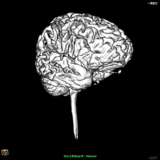
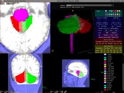
Gesamtes
Gehirn
Kleinhirn
mit Spline
Klinik für Neurologie
Universität Duisburg-Essen
Prof. Dr. D. Timmann-Braun
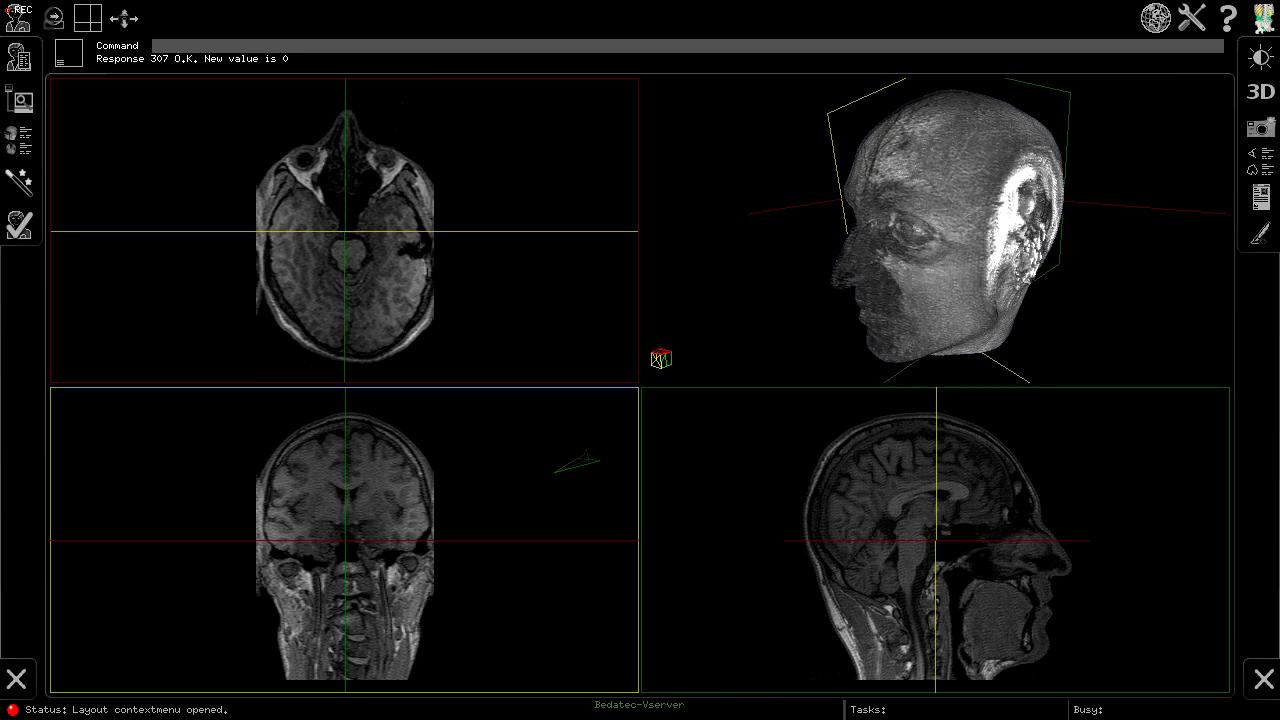
Dr. A. Dimitrova The MRI datasets were acquired at the Abteilung für Neuroradiologie des Instituts für Diagnostische und Interventionelle Radiologie der Universität Duisburg-Essen (Dr. E. Gizewski, Prof. Dr. M. Forsting). The volumetry is done using an ECCET-based application on ordinary PCs.
Kleinhirn
und
Hirnstamm
Challenges of Segmentation
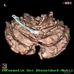
It is often very difficult to separate the cerebellum from neighbouring structures using only the grey values in the MRI datasets. There are many places where the grey values of brain and cerebellum touch without any visible grey value difference or separation. Simple thresholding is thus out of question to separate cerebrum from cerebellum. However we have developed sophisticated fill algorithms that often yield good results.
A human expert uses high level knowledge about the shape of the brain to correctly separate cerebrum from cerebellum. However it is not obvious how to transfer such intuitive knowledge into algorithms.
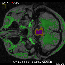
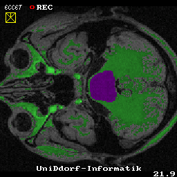
An even tougher problem is separating the cerebellum from the brain stem. There are large connections between them and it is a bit arbitrary where to draw the line. The image on the right shows all pixels that are within a given value range as green. The range is specified so that the brain stem is just included. Obviously, this also marks parts of the cerebellum.
Even dividing lines drawn by human experts vary with large inter- and intra-observer differences, so that it seems impossible to obtain a reliable way to quantify the cerebellar volume. It is thus important to find a method that derives such a dividing line in a reproducible way. Our method satisfies this criterion.
Untere Hüllenbegrenzung
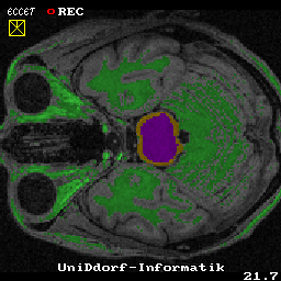
Obere Hüllenbegrenzung Some slices of the MRI data show no clear boundary between the cerebellum and the brain stem. The slices below and above however have such clear boundaries, which allows to manually or semi-automatically mark the brain stem cross section there.
Interpolierte Schicht Using an interpolation technique, the missing part is estimated from the shape of the marked cross sections. The result shows very few inter- and intra-observer differences.
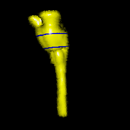
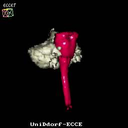
Hirnstamm
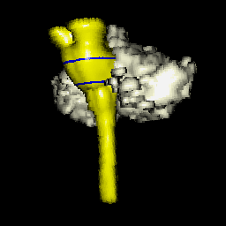
Hirnstamm The brain stem is shown in yellow on the pictures on the right. The blue lines mark the segmented slices between wchich the interpolation took place. The cerebellum is shown in white.